
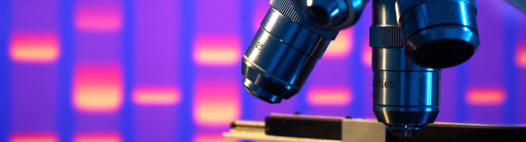
A microscope allows you to see incredibly detail in the tiniest of specimens. Surely they must all be considered, equal, right? In actuality there are two main types, namely the light microscope vs electron microscope.
Most likely you are familiar with a light microscope. This version sits on a desk, uses light and a lens to magnify your objects, and is often found in science classes around the world. However, when you need to see even more detail, particularly in a professional setting, an electron microscope is the tool you need to turn to. If you’re becoming interested in the world of science and want to understand how the two microscopes function, this article is for you. We’ll break down the different parts of the two microscopes and discuss their features and abilities. Our aim is to illuminate you on all the possibilities the light and the electron microscope bring.
If you’re completely new to the world of microscopes, a light microscope is the best place to start. It is the most common tool for science classes and props on TV shows. Even within this category, however, there are different types of light microscopes. For example, compound microscopes use two lenses for a better magnification process. Then there are digital microscopes which uses a digital camera for viewing, instead of an eyepiece.
There are both simple and complex parts that make up a
light microscope
Trusted Source
Microscopes. National Geographic Society
A microscope is an instrument that can be used to observe small objects, even cells.
www.nationalgeographic.org
, so we’ll do our best to explain them all.
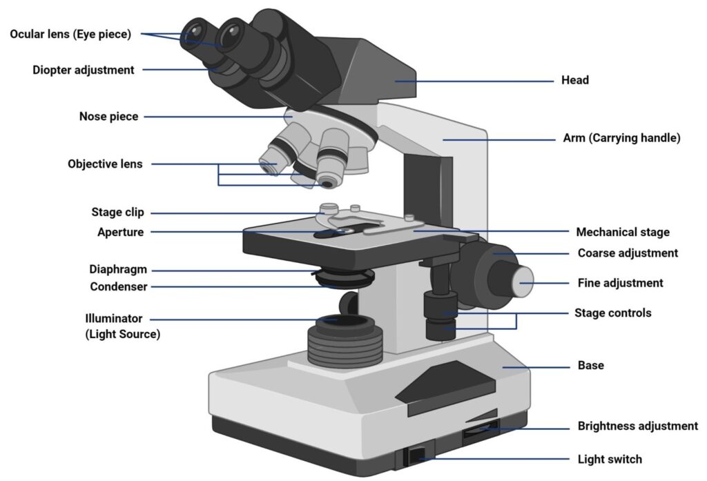
At the top of a light microscope, you’ll find the eye piece that you look through. This leads to the body tube which connects to the objective. There is a gap between the objective and the condenser, which is where you place your specimen. At the bottom of this area is a mirror base, which adds a magnification element. Finally, on the side of a light microscope are various knobs that allow you to adjust and focus.
A light microscope uses light and lenses to magnify an object. You can adjust the base for a thicker specimen although the use of thin slides is more commonly used.
Furthermore, because this telescope only uses light, you can view both alive and dead specimens. As a result, you can study live organisms such as bacteria and cells.
Those working in a professional lab will be more accustomed to an electron microscope. It is a very powerful tool that can actually allow you to see such tiny specimens, including electrons.
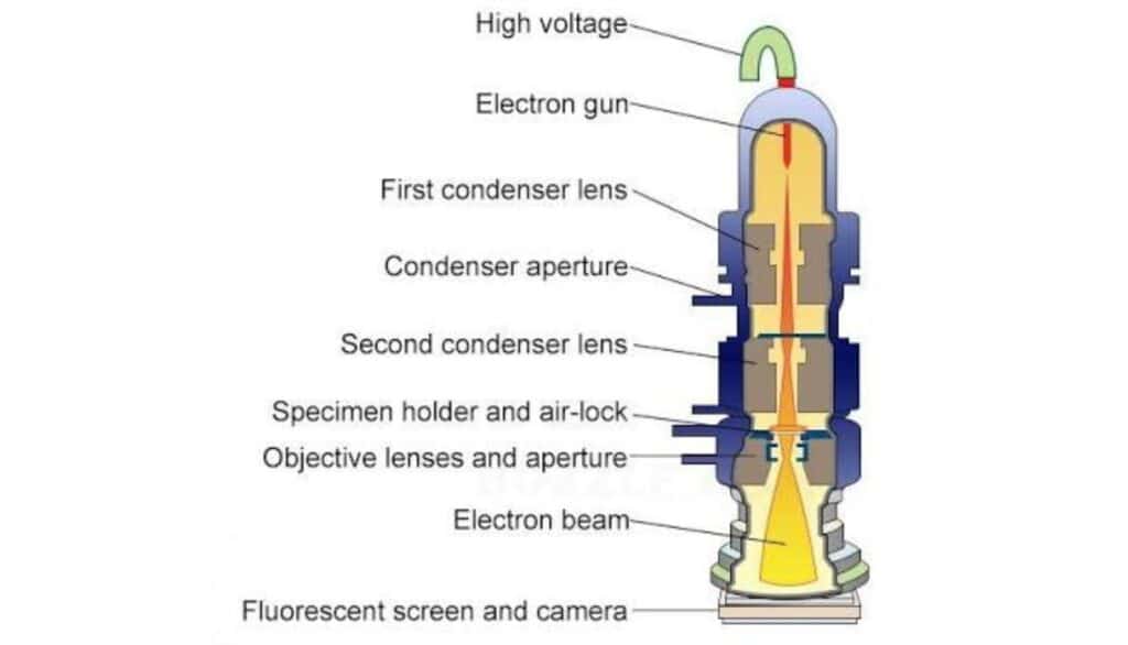
When thinking about an electron microscope, think about one large tube. At the top is an area with high voltage and the middle has lenses and apertures. Finally, at the bottom is an electron beam. You don’t have anywhere to look into as the image is then transported to a screen for viewing.
This might get a bit complicated, so we’ll do our best to offer a simple explanation. While a light electron simply uses light to magnify an object, an electron microscope uses electrons that are then passed through a specimen.
If you think of an electromagnetic spectrum, visible light is in the middle. However, there are many other parts to the spectrum, including ultraviolet, X-rays, and gamma rays, which is the area that the electron microscope operates on.
Once these rays pass through a specimen, they are projected onto a screen which is then magnified extremely powerfully, up to 2,000,000 times.
Let’s look at this handy chart to see how the two microscopes break down.
| Light | Electron | |
| Illuminating source | Light | Electrons |
| Max magnification | 1,000x | 2,000,000x |
| Specimen type | Live or dead | Dead or dried |
| Lenses material | Glass | Electromagnetic |
| Resolving power | 0.25µm-0.3µm | 0.001µm |
| Image | Colored | Black and white |
| Field of use | Study of internal structure | Study of external surface, cell structure and small organisms |
Perhaps the most distinct difference between the two microscopes is the view capability. While a light microscope does pretty well at 1,000x, an electron is incredibly more powerful, at up to 2,000,000x. That’s almost too hard to comprehend.
If you want a powerful microscope with a better magnification, you can look at a model such as the AmScope B120C. It has the ability of up to 2,500x and yet is actually quite affordable.
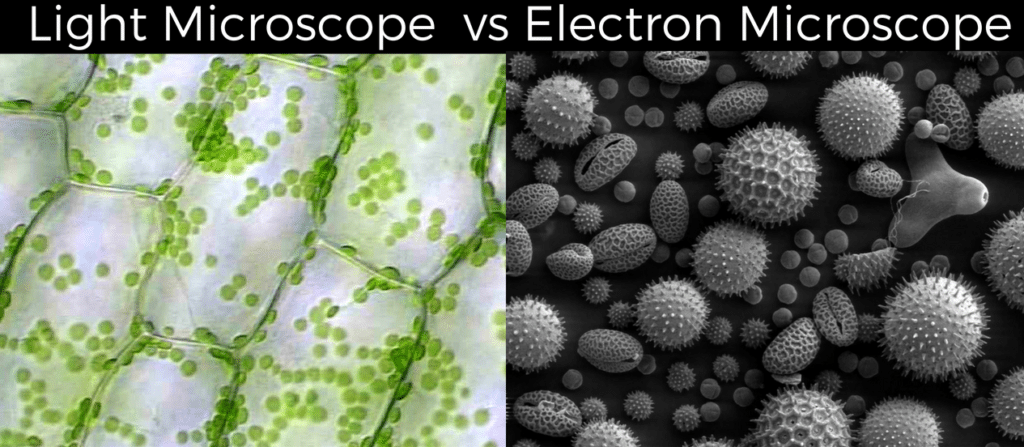
Another difference is how the specimen is views. With a light microscope, you only see the specimen in 2D. However, with an electron microscope, you can view it in 3D. The exception is with stereo microscopes, which uses two eyepieces to create a 3D image.
Finally, a light microscope allows you to see the specimen exactly how it is, meaning in full color. With an electron microscope, the image is seen in black and white. The good news is that a computer model can add color for a more realistic view.
As you can probably imagine, a light microscope is used for more simple study. It can be used in the classroom or the lab and is pretty versatile. You can see the internal structure of organisms and get a good sense of what makes up a specimen.
With an electron microscope, your field of use is much more specialized. It won’t be found in a high school setting, but instead in a high-tech facility. It is used to study cell structure, external surfaces, and microorganisms.
If you’ve read any news articles in the past year, chances are you’ve seen images of the coronavirus Trusted Source New Images of Novel Coronavirus SARS-CoV-2 Now Available | NIH: National Institute of Allergy and Infectious Diseases NIAID has produced images of the novel coronavirus SARS-CoV-2 on its scanning and transmission electron microscopes on Tuesday, Feb. 11, 2020. SARS-CoV-2 causes COVID-2019 disease, which has grown to be a global public health emergency since cases were first detected in Wuhan, China, in December 2019. www.niaid.nih.gov . These were most likely taken with the help of an electron microscope.
In simple terms, light microscopes use light, which is fairly harmless, while electron microscopes put off some ration, which is less-so. In fact, electron microscopes need to be registered with sate environmental regulators.
The good news is the construction of electron microscopes puts safety first. Still, you should always report any cracks or unusual deficiencies with an electron microscope.
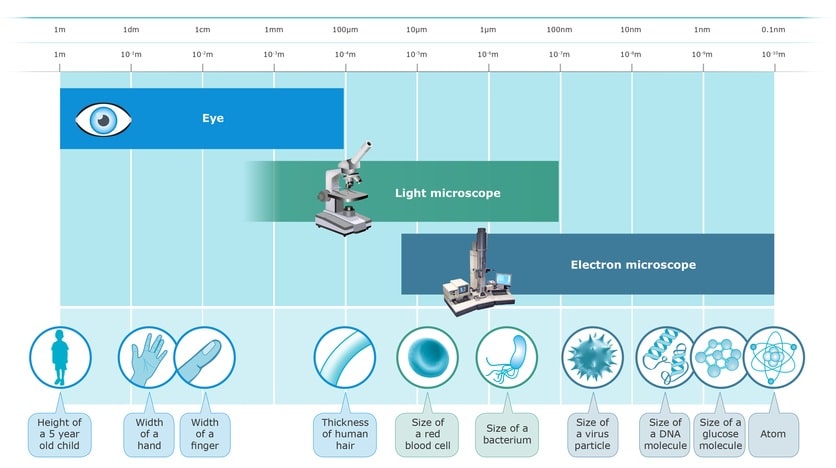
When discussing types of specimens used, it’s all about size. Larger specimens can be views with a light microscope while smaller, more intricate specimens need the use of an electron microscope.
For example, when performing biopsy examinations or even looking at the cells of crystals, an electron microscope is necessary. If you are looking at the skin of an onion for a science project, a light microscope will work just fine.
While the price range of light microscopes definitely varies, with some basic models being so affordable they can be used at home, electron microscopes are definitely not a piece of equipment you can simply pick up.
If you have a child who loves science, a product like the SWIFT Microscope SW150 is a great option. It is affordable yet powerful, with the ability to magnify up to 1000x.
Electron microscopes are only to be used by large companies that have the means to purchase them. Furthermore, you need to source them from a specific company, and not just your local electronics store.
Even though a light microscope is relatively heavy, it can be easily moved around. It will normally have a safety bar so you can attach it to a table to keep it steady and minimize the risk of it falling off
On the other hand, an electron microscope is definitely not portable. They are assembled in a lab and remain their permanently.
Even though light microscopes lack the view magnification ability of electron microscopes, they are still pretty versatile. You can collect samples from your garden, such as leaves and bugs, and inspire young scientists. You can also prepare slides of larger bacteria and see them up close.
In contrast, the immense viewing power of electron microscopes makes its versatility a lot narrower in focus. With this technology, you want to use it for microorganisms and cell research.
Whether you’re new to the area of science or just want to learn more, understanding the amazing tool that is a microscope is key. Without it, we just can’t see all the amazing parts of the world around us. Most are familiar with a light microscope, which can sit on a table and allow use to see objects in extremely vivid detail. We can see the veins in a leaf, the movement of large bacteria, and the layers of food. But if you want to go even further, the use of an electron microscope is needed. These have amazing magnification powers but their hefty size and price tag put them out of reach of most people. If you are a professional scientist, you may have access to an electron microscope but amateur enthusiasts will have to settle for the more affordable and convenient light microscope. We hope you now have a better understanding of the differences between a light microscope vs electron microscope. Just remember that the world around is quite large and sometimes the most important parts of the world can only be seen through a microscope.


 About us
  Contact Us